45 label the transmission electron micrograph of the nucleus
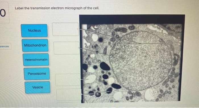
Solved Label the transmission electron micrograph of the - Chegg Expert Answer. 100% (4 ratings) Explanation - Mitochondrion is filamentous or globular in shape, occur in variable numbers from a few hundred to few thousands in different cells. It …. View the full answer. Transcribed image text: Label the transmission electron micrograph of the mitochondrion. Matrix granule Mitochondrion Outer membrane ... Solved Label the transmission electron micrograph of the Transcribed image text: Label the transmission electron micrograph of the cell. 0 Nucleus rences Mitochondrion Heterochromatin Peroxisome Vesicle ULAR bumit Click and drag each label into the correct category to indicate whether it pertains to the cytoplasm or the plasma membrane. ICF Contacts the ECF Made of proteins and lipids Surrounds the cell Contains lon channels Organelles Fibers, tubulos, passages, and compartments Cytoskeleton Plasma Membrane Cytoplasm Correctly identify this tissue ...
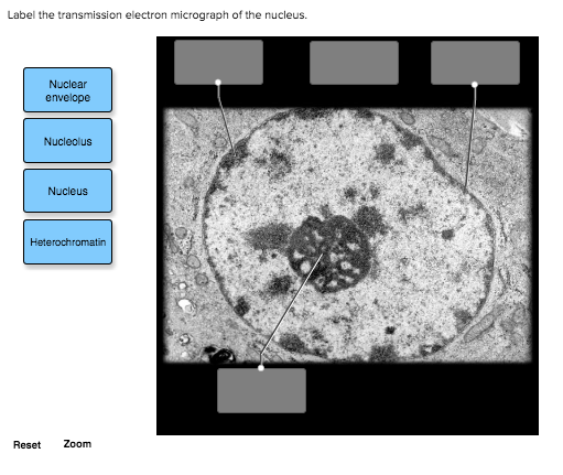
Solved Label the transmission electron micrograph of the - Chegg Expert Answer. 100% (23 ratings) Transcribed image text: Label the transmission electron micrograph of the nucleus. Nuclear envelope Nucleolus Nucleus Heterochromatin Reset Zoom.
Label the transmission electron micrograph of the nucleus
a Transmission electron micrograph of leaf cells from E. osiris. Plant ... Labels: Ch, chloroplast; Ne, nucleus; V, vacuole. b Transmission electron micrograph of root cells from E. osiris. Plant was exposed to 0 mg L⁻¹ (control, c, e, g × 30,000) and 15 mg L⁻¹(d ... Electron Micrographs Note: When comparing sizes from one micrograph to another, remember to consider the respective magnifications. Figure 1 Micrograph of a nucleus. 1. Labeling the Cell Flashcards | Quizlet Label the transmission electron micrograph of the nucleus. membrane bound organelles golgi apparatus, mitochondrion, lysosome, peroxisome, rough endoplasmic reticulum nonmembrane bound organelles ribosomes, centrosome, proteasomes cytoskeleton includes microfilaments, intermediate filaments, microtubules Identify the highlighted structures
Label the transmission electron micrograph of the nucleus. To label: The transmission electron micrograph of a relaxed sarcomere ... Start your trial now! First week only $4.99! arrow_forward Literature guides Concept explainers Writing guide Popular textbooks Popular high school textbooks Popular Q&A Business Accounting Economics Finance Leadership Management Marketing Operations Management Engineering Bioengineering Chemical Engineering Civil Engineering Computer Engineering Computer Science Electrical Engineering ... Label This Transmission Electron Micrograph Of A Relaxed ... - Blogger Label this transmission electron micrograph of relaxed sarcomeres by clicking and dragging the labels to the correct location . Label the following image using the terms provided. Note how the sarcomeres are extended to only approximately 120 % . IMG_2132 - FIGURES Label this transmission electron from Transmission electron micrograph of rectal epithelial cell showing ... Download scientific diagram | Transmission electron micrograph of rectal epithelial cell showing nucleus (N) with nucleolus (NU) and lysosomes (LY). 3400. from publication: Epithelium of the ... Transmission Electron Microscope (With Diagram) - Biology Discussion The final image in a TEM is known as transmission electron micrograph. The salts of some heavy metals, e.g., lead; osmium, tungsten and uranium are often used for staining. These heavy metal stains are used to increase the contrast between ultra structures and the background. The metals can be fixed on to the specimen and is referred to as ...
Label This Transmission Electron Micrograph - Kaiden Brown Label the transmission electron micrograph of the nucleus. Label the transmission electron micrograph of the nucleus. Transmission electron microscopy (tem) is a microscopy technique in which a beam of electrons is transmitted through a specimen to form an image. Figures label this transmission electron micrograph ( 16, . CIN2003. Ian Roberts. BIO 224: Lab Midterm Review Flashcards | Quizlet Loosely coiled fibers containing protein and DNA within the nucleus. false. T or F: The division of the cell's nuclear parts is called interphase. 46. ... Label the transmission electron micrograph of the mitochondrion. Synthesizes protein for secretion, insertion into the plasma membrane, and lysosomal enzymes. ... Transmission Electron Micrograph Of A Cell Nucleus Transmission Electron Micrograph Of A Cell Nucleus is a photograph by Cnri/science Photo Library which was uploaded on September 24th, 2018. The photograph may be purchased as wall art, home decor, apparel, phone cases, greeting cards, and more. All products are produced on-demand and shipped worldwide within 2 - 3 business days. Transmission electron micrograph of cell nucleus Transmission electron micrograph of the nucleus (circular) of a mouse liver cell. Surrounding the nucleus is delicate nuclear membrane, which contains gaps called nuclear pores that allow large molecules to pass out into the cell cytoplasm. The dark area in the lower part of the nucleus is the nucleolus.
Bio101 - Ch 6 HW Flashcards | Quizlet Study with Quizlet and memorize flashcards containing terms like Which of the following choices correctly matches a tool and its proper application? See Concept 6.1 -cell fractionation to study the function of specific organelles -light microscopy to study the internal structure of cilia -transmission electron microscopy (TEM) to study the surfaces of preserved cells -transmission electron ... Transmission Electron Micrograph of transfected HL-1 cells labeled for ... The label (large dot) is associated with the endoplasmic reticulum (arrows), nuclear envelope, and the nucleus (N). ... Transmission Electron Micrograph of transfected HL-1 cells labeled for ... A tour of the cell: View as single page - Open University Figure 2 (a) A transmission electron microscope. (b) A transmission electron micrograph of a frog leukocyte (white blood cell). The nucleus and nucleolus (Section 4.3), mitochondria (Section 4.10) and Golgi apparatus (Section 4.7) can be seen. The dark area of the nucleus contains densely packed DNA. Transmission electron micrograph (TEM) identifying immunogold labeled ... (B) Immunogold labeling of ESR1 identified the protein localization at the plasma membrane, the cytosol, and the nucleus. (C) Illustrates the location of Cav-1 close to or at the plasma membrane.
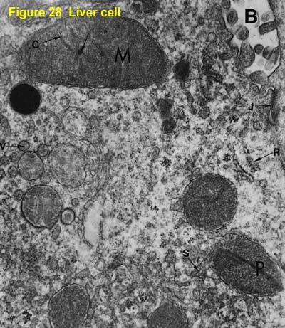
2.3.3 Identify structures from electron micrographs of liver cells
anatomy 10.png - Label the transmission electron ... anatomy 10.png - Label the transmission electron micrograph of the. anatomy 10.png - Label the transmission electron micrograph... School Utah Valley University; Course Title ZOOL 1090; Uploaded By emileeroylance19. Pages 1 Ratings 67% (3) 2 out of 3 people found this document helpful;
A tour of the cell: View as single page
Transmission Electron Micrograph Of A Cell Nucleus Art Print Transmission Electron Micrograph Of A Cell Nucleus Art Print by Cnri/science Photo Library. All prints are professionally printed, packaged, and shipped within 3 - 4 business days. Choose from multiple sizes and hundreds of frame and mat options.
Frontiers | Contributions of Ultrastructural Studies to the ...
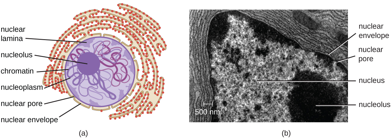
Cell Organelles Electron Micrograph Lab.pdf - Cell... Cell Organelles Electron Micrograph Lab Label Structure Form Function 1 Nucleus Contains the nuclear envelope and chromatin Stores the genetic material DNA, which governs the characteristics of the cell and its metabolic functioning 2 Nuclear envelope Surrounds around the nucleus, which contains genetic material protects all important genetic information from the chemical reactions that take place outside the nucleus 3 Chromatin Looks like beads on a string, beads=nucleosome Chromatin allows ...
A tour of the cell: View as single page
Electron micrograph of the anteroventral cochlear nucleus immunolabeled ... This micrograph illustrates the overall pattern of endbulb - synapse terminal profiles along the somal membrane. One of these terminals ( asterisk ) is shown at high magnification in the inset.

1.2 Skill: Interpretation of electron micrographs
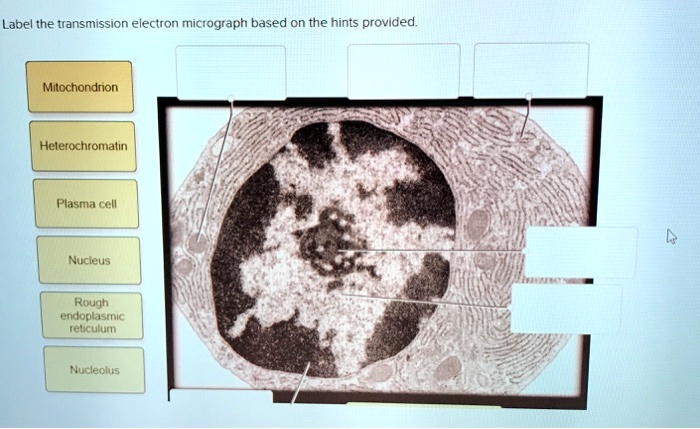
Study Chapter 14 & 15 Flashcards Flashcards | Quizlet Label the transmission electron micrograph based on the hints provided. Place the following tonsils in order based on their location from superior to inferior. Label the structures in the photomicrograph based on the hints provided.

Electron micrograph of a rabbit retina grown in situ (p"14 ...
Label the transmission electron micrograph of the nucleus. - Transtutors Label the transmission electron micrograph of the cell. 0 Nucleus rences Mitochondrion Heterochromatin Peroxisome Vesicle ULAR bumit Click and drag each label into the correct category to indicate whether it pertains to the cytoplasm or the plasma...

TEM images of cell organelles such as the nucleus and ...
PDF Identifying Organelles from an Electron Micrograph Courtesy of Dr. Julian Thorpe - EM & FACS Lab, Biological Sciences University Of Sussex. A single Granum Chloroplast envelope visible as two membranes Stroma containing numerous small ribosomes Lamellae connecting different grana. Lipid droplets. Title.
SOLVED: Label the transmission electron ricrograph based on ...
Transmission electron microscopy (TEM) imaging of ... Download scientific diagram | Transmission electron microscopy (TEM) imaging of nuclear envelope and nucleoplasmic reticulum (NR) in salivary gland nuclei.
IJMS | Free Full-Text | Visualization of Chromatin in the ...
Label This Transmission Electron Micrograph : The Corresponds To The ... Label the transmission electron micrograph of the. Transmission electron microscopy (tem) is a microscopy technique in which a beam of electrons is transmitted through a specimen to form an image. Subset of labeled images and transfer labels to the entire image corpus. Provide the labels for the electron micrograph in figure 12.8. No microtubule labeling is evident.
Solved Label the transmission electron micrograph of the ...
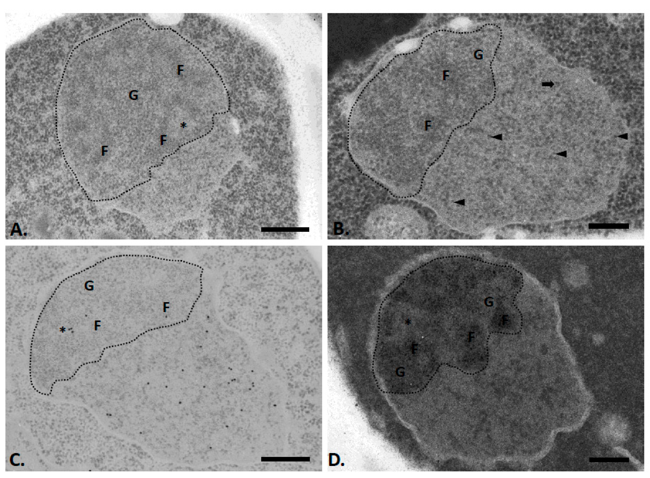
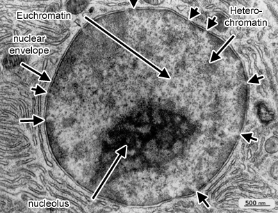
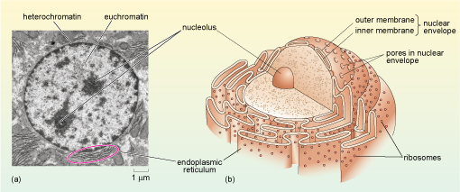
Electron Micrographs - University of Oklahoma Health Sciences Center Electron Micrographs** Below is a collection of electron micrographs with labelled subcellular structures that you should be able to identify. Also, be sure to observe any electron micrographs which are made available in the laboratory by the instructor. ... Figure 1 Micrograph of a nucleus. 1. Heterochromatin 2. Euchromatin 3. Nucleolus 4 ...
A tour of the cell: View as single page
A&P Unit 2 Exam Flashcards | Quizlet The complement system Label the transmission electron micrograph based on the hints provided. Plasma cells produce antibody molecules. Name the cells included in the mononuclear phagocytic system. macrophages monocytes neutrophils An immunoglobulin molecule is an antigen secreted by T lymphocytes. False trabecula of spleen thoracic duct
Solved Label the transmission electron micrograph of the ...
Labeling the Cell Flashcards | Quizlet Label the transmission electron micrograph of the nucleus. membrane bound organelles golgi apparatus, mitochondrion, lysosome, peroxisome, rough endoplasmic reticulum nonmembrane bound organelles ribosomes, centrosome, proteasomes cytoskeleton includes microfilaments, intermediate filaments, microtubules Identify the highlighted structures
The Cell: The Histology Guide
Electron Micrographs Note: When comparing sizes from one micrograph to another, remember to consider the respective magnifications. Figure 1 Micrograph of a nucleus. 1.

Electron micrographs of SPIO-labeled MSCs. A, Cell nucleus (N ...
a Transmission electron micrograph of leaf cells from E. osiris. Plant ... Labels: Ch, chloroplast; Ne, nucleus; V, vacuole. b Transmission electron micrograph of root cells from E. osiris. Plant was exposed to 0 mg L⁻¹ (control, c, e, g × 30,000) and 15 mg L⁻¹(d ...

Transmission electron micrographs of the follicle cell ...
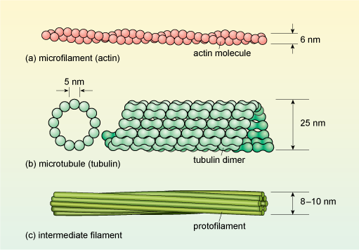
3.4 – Unique Characteristics of Eukaryotic Cells ...

Immunogold labelling - Wikipedia

Transmission electron micrographs of liver cells of the ...

anatomy 10.png - Label the transmission electron micrograph ...

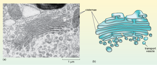
Biology 2e, The Cell, Cell Structure, The Endomembrane System ...

Lab2.docx - Lab assessment #5 Part A – Assessments 1. Label ...
Solved Label the transmission electron micrograph of the ...

False Colour Transmission Electron Microscope (tem ...
A-level Biology Bridging Course - Week 1
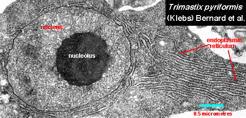
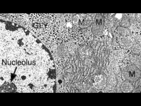
Nucleolus Electron Micrograph

Representative transmission electron micrographs of pancreas ...
A tour of the cell: View as single page

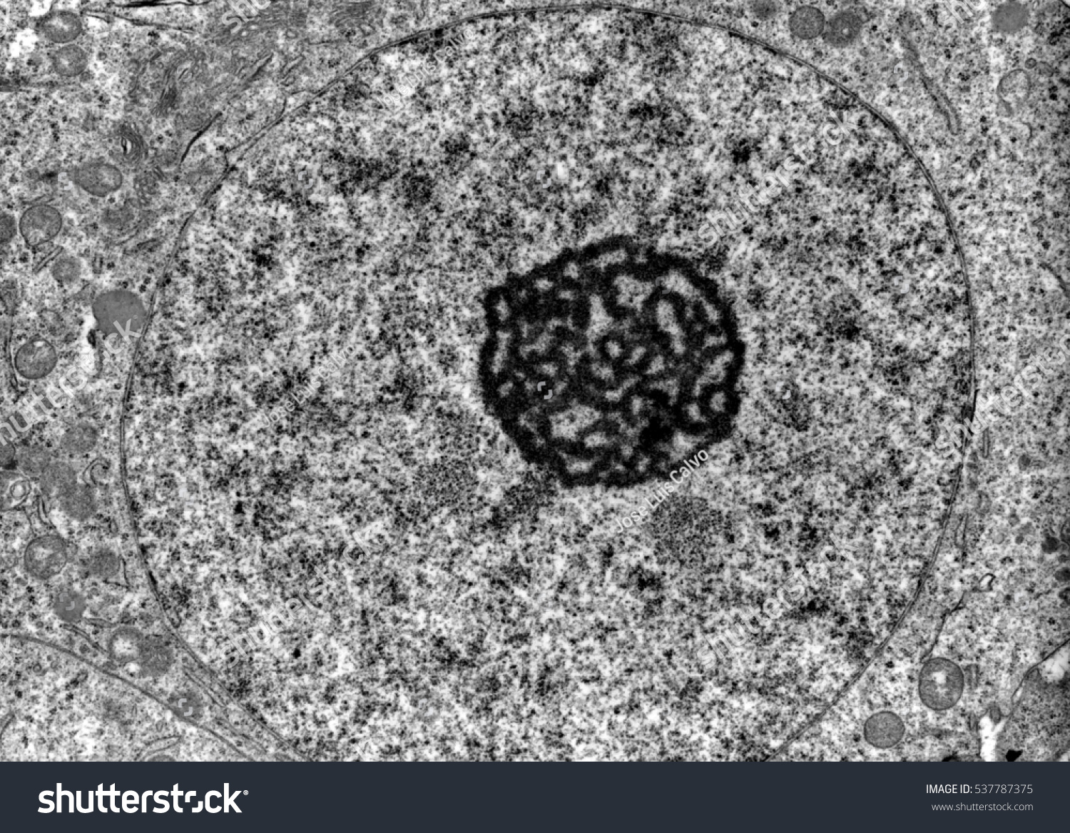
Identifying the Parts of the Nucleus in an Electron Micrograph
Transmission Electron Microscope Tem Micrograph Showing Stock ...

Biology: 1.2 Ultrastructure of Cells Flashcards | Quizlet
Electron Micrographs
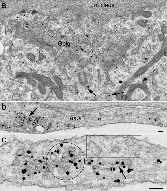
Immunogold labeling of synaptic vesicle proteins in ...
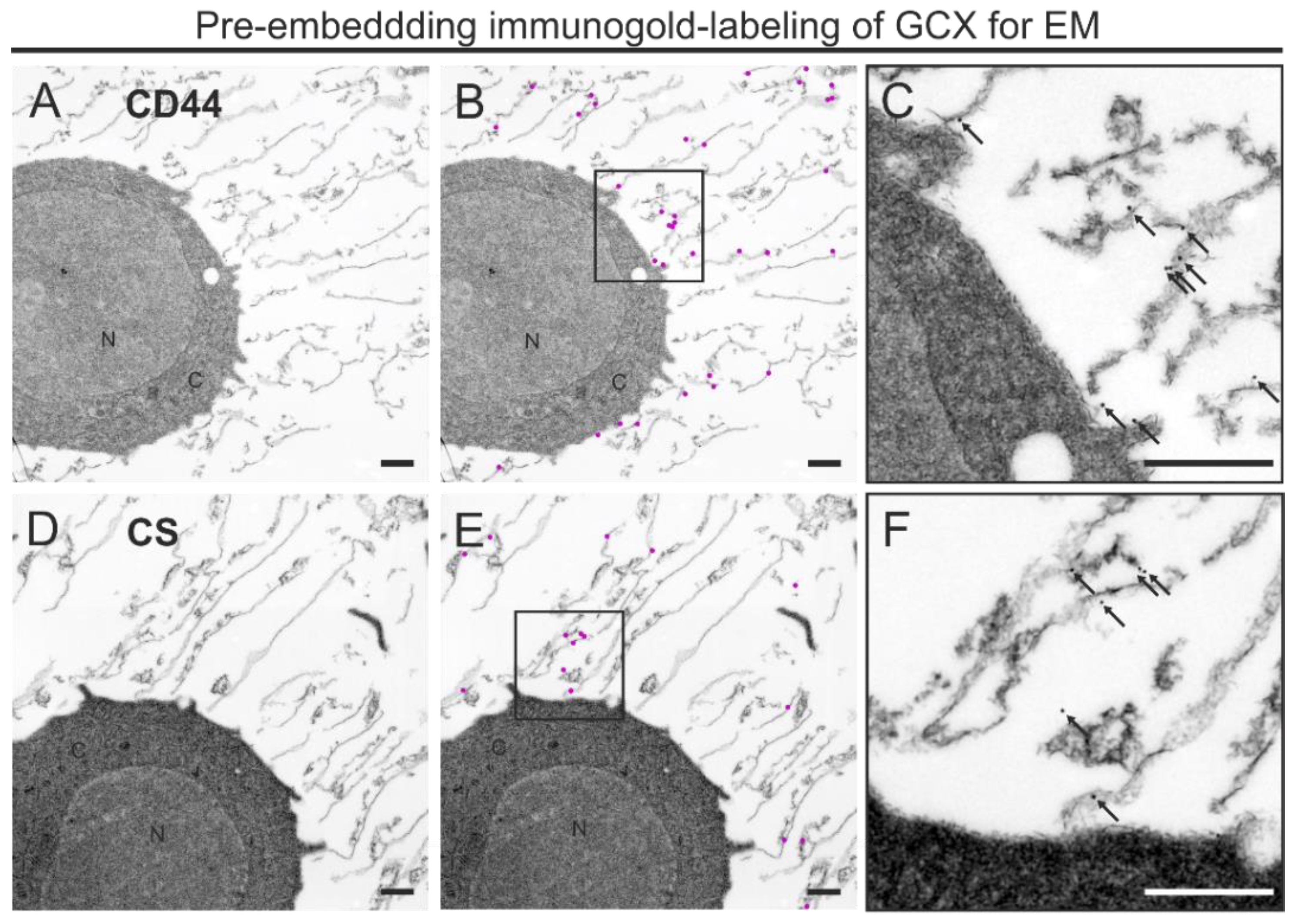
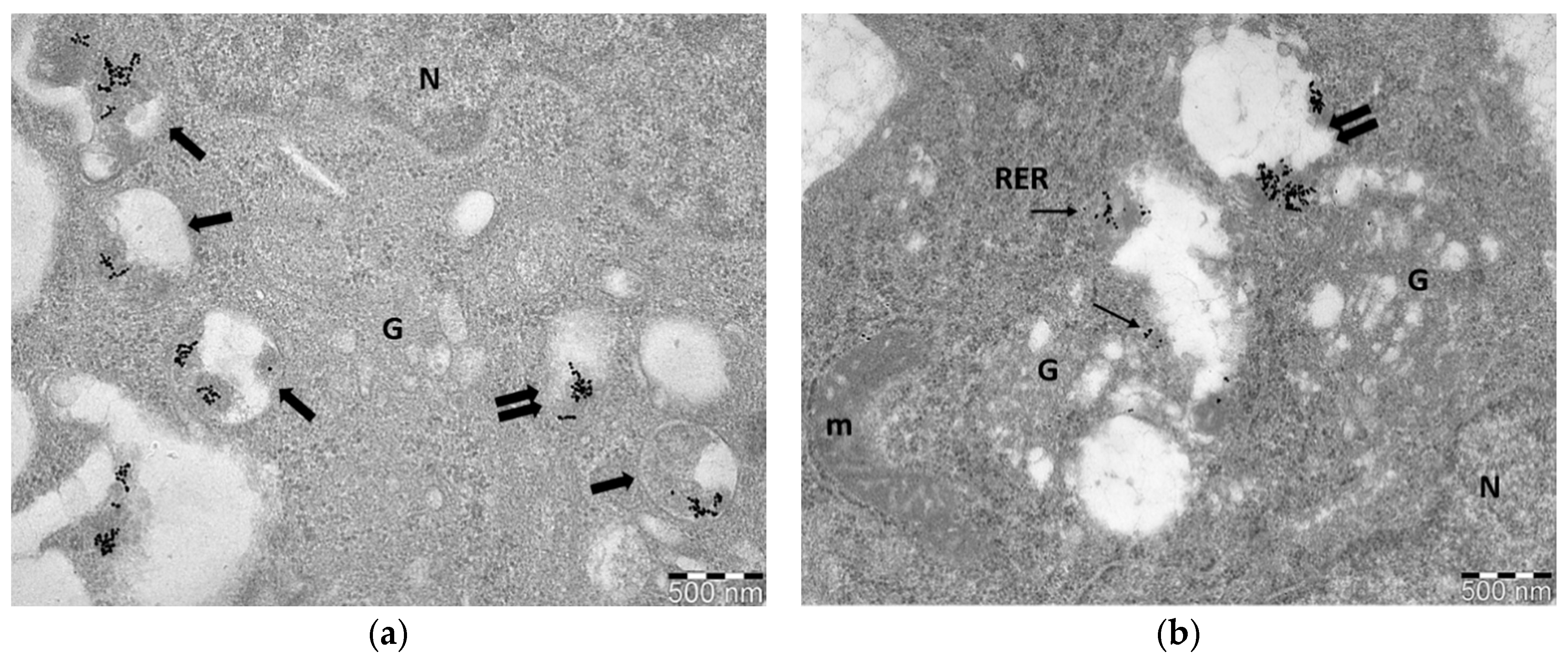
Biology | Free Full-Text | Immuno-Electron and Confocal Laser ...
What is a diagram of a plant and animal cell under an ...
Show Question
Solved] FIGURE 5.5 Transmission electron micrographs of ...

Transmission electron microscope (TEM) micrograph showing the ...

1.1 Cell structure | Cells as the basic units of life | Siyavula

Mitochondria | BioNinja

Transmission electron microscopy (TEM) imaging of nuclear ...
Transmission electron microscope (TEM) micrograph showing the ...
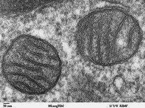
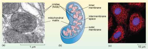
Mitochondrion - Wikipedia
Nanomaterials | Free Full-Text | A Guide for Using ...

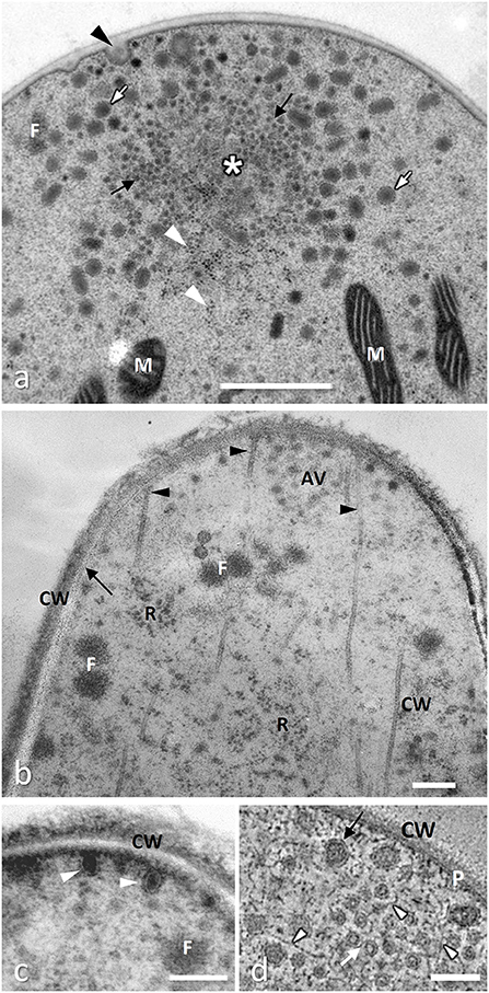
Sub-urothelium. Electron microscopy. (A) A fibroblast (FB ...
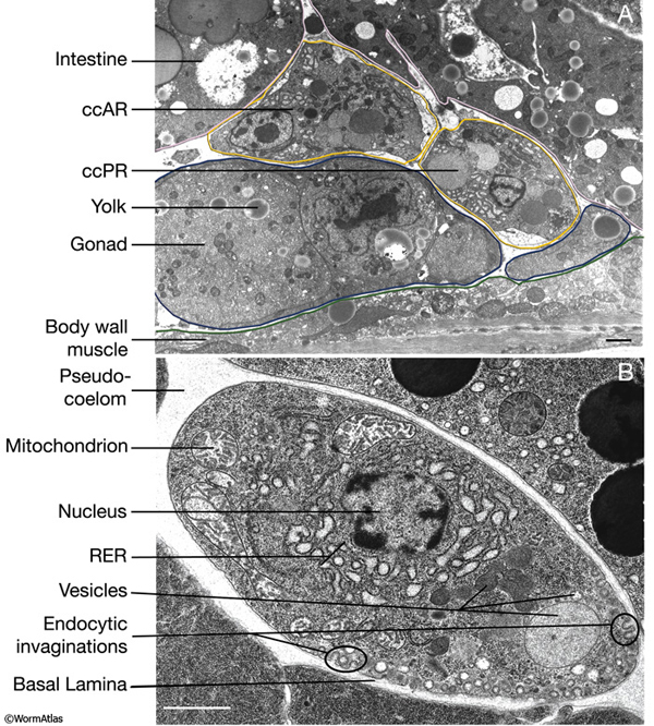
Hermaphrodite Coelomocyte System


Komentar
Posting Komentar